What is Insulin Resistance and What are Its Causes
For many of my patients, the diagnosis insulin resistance (IR) is the turning point that makes them turn to a nutritionist.
This is because the condition gradually progresses into prediabetes, metabolic syndrome, and type 2 diabetes (T2D).
Furthermore, IR is associated with an increased risk of polycystic ovaries, heart attacks, strokes, and even some types of cancer!
But a proper diagnosis requires a comprehensive assessment – symptoms, lab values, weight, lifestyle – not just a few lab values!
Therefore, it is important to discuss the results with experienced specialist – nutritionist or endocrinologist to avoid misdiagnoses and unnecessary stress.
In the case of IR, the experienced nutritionist will also suggest the most effective treatment method – diet therapy that is individualized, balanced, and sustainable enough for you to follow in the long term.
What is insulin resistance?
Insulin resistance is a condition in which cells in the body become less responsive to insulin signals. This happens because their insulin receptors are decreased sensitivity.
Insulin itself is an anabolic (building) hormone produced by the pancreas. [1].
Almost everyone knows that insulin works by „unlocking“ cells to take in glucose from the blood. This glucose is used for energy or stored:
- As glycogen: In muscles and liver (these are limited depots).
- As fat: When glycogen stores are full, excess glucose is converted to fat and stored in fat cells.
- As an anabolic hormone, insulin also prompts muscles to take up amino acids, which is key to muscle growth.
But insulin also has very important suppressive effects!
Much of the vicious cycle that occurs with IR is due to the fact that some of the effects of insulin are ineffective in IR.
In a person without IR, insulin has extremely important functions for:
- Suppression of the production of new CD in the liver
- Suppression of glycogen and protein breakdown in muscles
- Stops the release of free fatty acids (FFA) from adipose tissue
Insulin resistance develops when the cells of these three key tissues – muscle, liver and fat tissue – lose their insulin sensitivity!
This disrupts all three of the above-mentioned functions of insulin in the relevant tissues.
Why do I have insulin resistance? Where does the development of insulin resistance begin?
The development of insulin resistance most likely does not begin simultaneously in the liver, muscle, and adipose tissue.
There are myths that with insulin resistance, muscle tissue becomes more resistant than fat, and this directs more insulin to fat stores.
This is false, because in fact, adipose tissue also becomes insulin resistant during IR, and it is even very likely that the problem starts there.
In this case, insulin cannot do its job in fat depots - to suppress the uncontrolled release of free fatty acids (FFA) into the bloodstream.
These SMCs can enter other tissues and organs, such as muscle tissue.
There, the SMCs are converted into metabolic intermediates that damage the insulin resistance of the cells.
Their accumulation in the muscles is detrimental to the normal function of insulin, since in healthy people around 80% from the circuit breaker is consumed by skeletal muscles [2].
The liver usually develops IR as a consequence of resistance from muscle and fat depots.
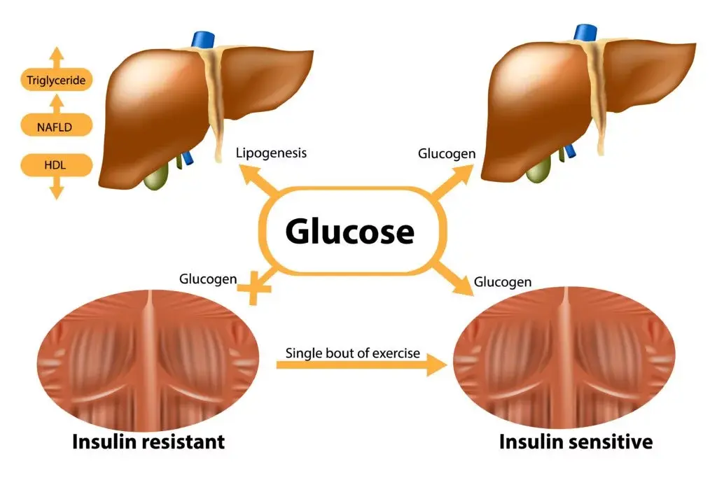
- When muscles are resistant, this directs the flow of carbohydrates to the liver, which converts them into fat and becomes fatty.
- Similarly, IR of adipose tissue directs the flow of BMC to the liver, which again makes it fatty and resistant.
Liver resistance prevents insulin from effectively suppressing the production of new glucose, which leads to a gradual increase in blood sugar levels and progression to prediabetes and type 2 diabetes.
High insulin also increases the risk of polycystic ovaries in women.
What is the difference between diabetes and insulin resistance?
A gradual rise in blood sugar does not immediately lead to a diagnosis of diabetes. In fact, fasting glucose should be below 5.6 mmol/L, and a diagnosis of diabetes is made with a level above 7 mmol/L. When blood sugar is between these two values, it is called prediabetes.
Prediabetes is a sure sign of IR.
It is often accompanied and even preceded from high insulin, abdominal fat accumulation, abdominal obesity, high blood pressure, poor lipid profile, high androgens in women and low testosterone in men.
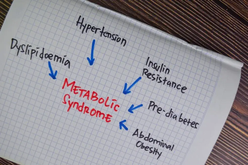
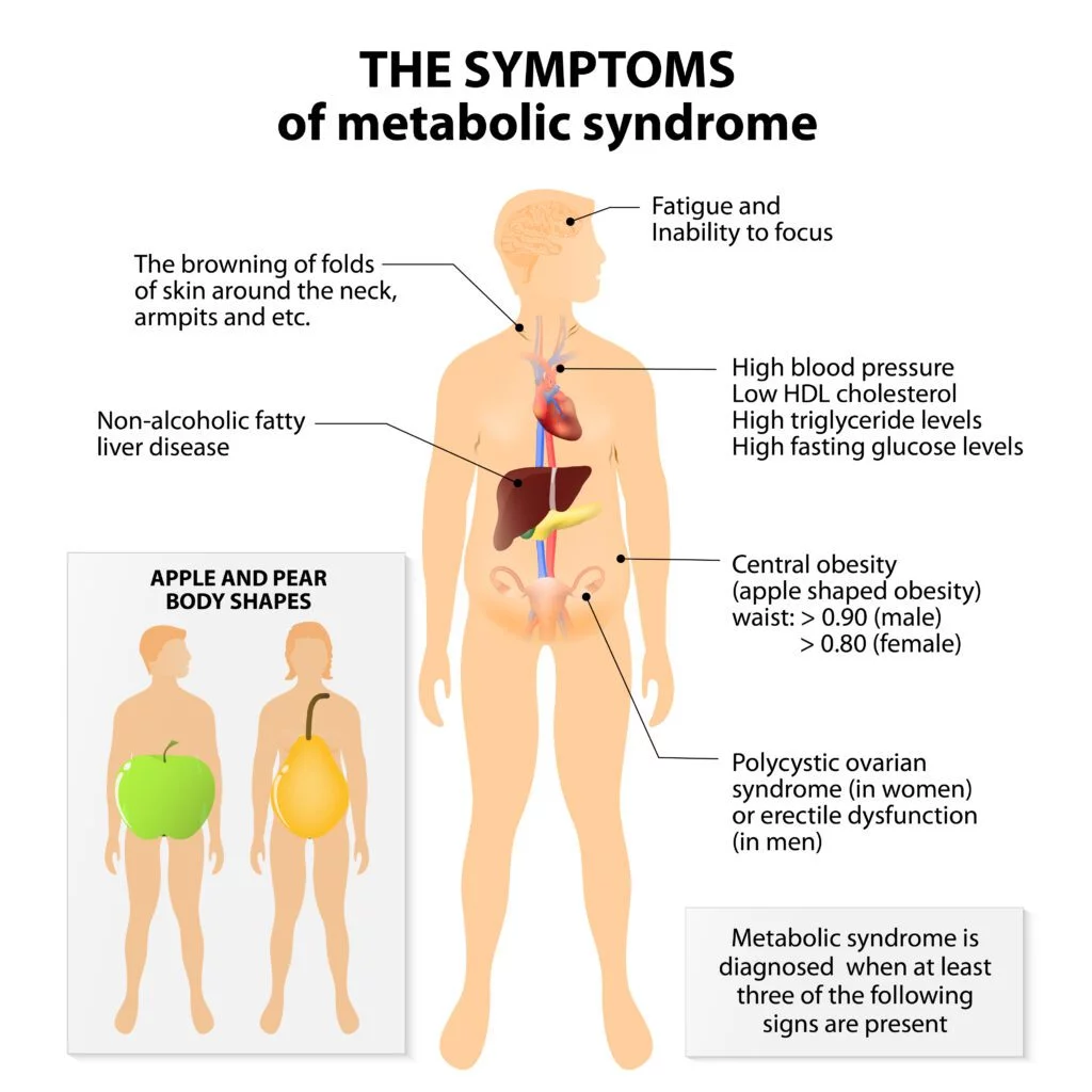
If at least three of the following symptoms are present, then we can also talk about insulin resistance syndrome, or metabolic syndrome: [3],[4]:
- Pre-diabetes (fasting BMI over 5.6)
- High triglycerides (over 1.7 mmol/l)
- Low HDL „good“ cholesterol (below 1.3 mmol/L)
- High blood pressure (over 130/85)
- Abdominal circumference over 80cm for women or 94cm for men (more precisely, the ratio is over 0.5 for waist/height)
Diacylglycerols can also accumulate in the pancreas, where they likely reduce its capacity to produce insulin.
The combination of reduced capacity and insulin levels, with increased needs, leads to an increase in BG above 7 and the condition progresses to type 2 diabetes.
Statistics show that over 501% of people with IR progress to stage 2. [5].
The good news is that IR and even incipient type 2 diabetes mellitus are considered reversible states, but only through timely and decisive lifestyle changes.
Is insulin to blame for the insulin resistance epidemic?
The insulin sensitivity of the three main tissues – muscle, fat, and liver – may be reduced for two main reasons – a reduced number of insulin receptors, or suppression of the signaling of the receptors themselves.
Most people imagine that excess insulin and glucose causes decreased sensitivity by reducing the number of receptors.
But in fact, long before that, the signaling of the receptors themselves is suppressed, and insulin or glucose have nothing to do with it.
As mentioned, this damage is most likely due to the accumulation of substances like diacylglycerols. These accumulate due to many factors, such as excess calories, increased inflammation, various hormones, and more.
They accumulate in the membranes of muscle cells, liver, and adipose tissue. The development of IR in these tissues leads to an increase in serum insulin, not the other way around.
It is actually quite normal for insulin secretion to be stimulated when blood glucose and/or amino acid levels (from protein in food) increase.
Furthermore, insulin secretion never stops in healthy people. The pancreas maintains a constant, basal level of insulin even during fasting.
Because insulin stimulates fat storage, it is often blamed for the obesity epidemic, which in turn is linked to IR.
There is a belief that maintaining low insulin protects against weight gain and prevents the development or progression of IR.
But, the truth is that fat accumulation depends on total calorie balance, and not just from insulin:
- If you are in calorie deficit (you take in fewer calories than you burn), you will lose weight despite insulin peaks after each meal.
- If you are in calorie surplus (you take in more calories), you will gain weight, even if your insulin is relatively low (it is never 0) and without peaks.
The body can store fat even when insulin is low.
For example, when you eat fat, another hormone called acylation-stimulating protein (ASP), which directs excess fat directly to fat depots [6].
That's why there are people who don't consume carbohydrates and still can't lose weight - they are not in a calorie deficit and fat is accumulated through ASP.

What causes insulin resistance?
IR is the result of the accumulation of several types of stress on the body – energy excess, genetic predisposition, lipotoxicity, lack of movement and muscle loss, and even the use of certain medications.
Genetic predisposition
Insulin resistance has a clear genetic predisposition [7]. However, there is no specific gene that fully explains IR. The condition has a polygenic basis, as dozens of genetic variants have been found that [8],[9]:
- affect insulin signaling and glucose metabolism (IRS1, GCKR, GRB14)
- regulate adipose tissue distribution and adipose tissue insulin sensitivity (PPARγ, NAT2)
- affect appetite and risk of obesity (FTO, MC4R)
- affect both insulin sensitivity and β-cell function (TCF7L2).
However, large clinical trials show that lifestyle changes can significantly reduce or completely avoid the risk of IR and type 2 diabetes – regardless of genes. [10].
Genes set the predisposition, but do not guarantee disease. Energy balance, physical activity, and weight often have a stronger effect than genetic predisposition.
Chronic energy surplus
Studies show that fat accumulation, even within normal body weight, can lead to the development of insulin resistance. [11], [12].
For example, one study on the subject shows that if you overeat for just one month and gain about 3 kg, it can reduce your insulin sensitivity by over 10 %, as measured by [13]:
- increased fasting insulin and glucose levels;
- higher HOMA-IR;
- decreased insulin sensitivity, measured with the gold standard – hyperinsulinemic euglycemic clamp.
Another study in young, weak and physically active people show that just one day of binge eating (with 78% over the required calories, mostly fat) leads to 28% decline in overall insulin sensitivity [14].
In this case, this decrease was measured by the change in the blood glucose curve after OGTT, while fasting indicators remained unchanged.
The likely cause is the rapid accumulation of lipids in the muscles and liver. This activates specific enzymes (PKC-θ and PKC-ε) that block insulin signaling at the cellular level.
Another study shows that weight gain can lead to insulin resistance even in children in about 1/3 of cases.rd of the cases [15].
And if there is already advanced obesity, a study of 3,423 people shows that the likelihood of IR is about 32 times greater than in people of normal weight. [16].
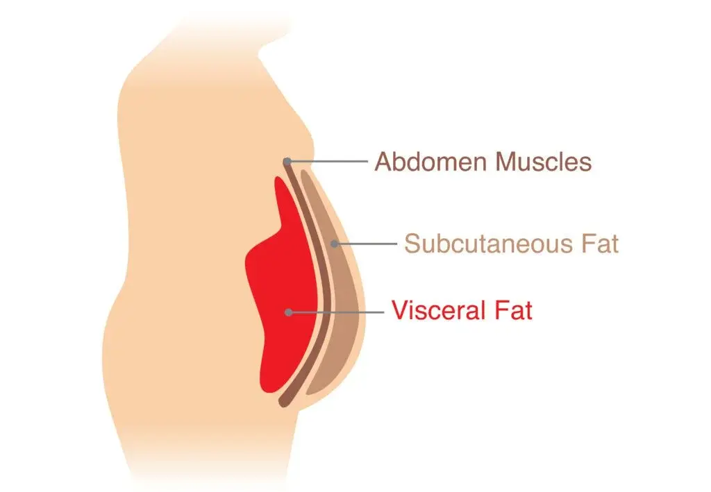
Obesity with accumulation of visceral and ectopic fat
When the cells of the subcutaneous adipose tissue reach the limit of their expansion, as well as due to hormonal or other reasons, the excess energy is forcibly redirected, in the form of free fatty acids (FFA), to:
- Fat located around the organs, called visceral [17]
- Fat in some organs, such as the liver and muscles, which is called ectopic
Multiple studies confirm that high levels of SMC in the blood are associated with IR [18], [19], [20].
This leads to IR of the three key tissues regulating insulin sensitivity [21], [22], [23].
It is difficult to say which tissue is affected first, but the involvement of adipose tissue is expressed in the following:
- Under stress and reduced blood circulation due to fat overload, adipose tissue develops inflammation and reduces its ability to respond to insulin.
- Without insulin, the synthesis of glycerol-3-phosphate, which normally keeps fatty acids inside in the form of triglycerides, is disrupted.
- At the same time, due to IR, insulin can no longer suppress lipolysis. The result is a massive release of SMC.
- Some of them are redirected to visceral fat depots, but they develop IR even faster, and lose BMC even more easily.
The following processes develop in parallel in the liver:
- Ectopic fats accumulate in the liver – lipid metabolites, especially diacylglycerol (DAG).
- DAG activates enzymes (PKC-ε) that block insulin signaling, which is why insulin is not suppressed by the liver and it does not stop producing glucose.
- Paradoxically, glucose continues to freely enter the liver through the insulin-independent transporter GLUT2 and it continues to synthesize new SMCs.
The following happens in muscle tissue:
- Ectopic fat (DAG) infiltration into the muscle activates an enzyme similar to that found in the liver (PKC-θ). This again sabotages insulin signaling and blocks the export of GLUT4 to the membrane.
- Thus, the muscles are left without energy, and the unabsorbed glucose returns to the liver and is transformed into BMC.
Immobilization, aging and loss of muscle tissue
A sedentary lifestyle also reduces insulin sensitivity. [24], [25].
Muscle absorbs glucose not only under the influence of insulin, but also directly during contraction.
During physical activity, its transporter – GLUT4 – increases, and glucose is absorbed simultaneously and without the need for insulin.
Conversely, with a sedentary lifestyle:
- large muscle groups hardly contract
- GLUT4 expression and mobilization decreases
- in practice, a large part of the muscles becomes an inefficient organ for absorbing glucose
When a sedentary lifestyle continues for years, and especially if it is accompanied by irregular eating and irregular or low protein intake:
- muscle mass, especially in the limbs, decreases (sarcopenia);
- the total reservoir in which glucose can be stored after a meal also decreases;
- sarcopenic obesity develops, in which the waist is increased even with normal or only slightly overweight weight
Low physical activity also accelerates muscle aging, leading to:
- Mitochondrial dysfunction and oxidative stress
- Myosteatosis develops (ectopic fat in the muscles, most often DAG) in which IR develops even with a normal amount of muscle mass.
All of this suppresses normal insulin signaling and leads to IR of both muscle tissue and the whole body.
But studies in older people who exercise regularly and maintain good physical fitness, insulin sensitivity and mitochondrial function may be similar to those of younger, more trained individuals. [26], [27].
That is, much of the "age-related" IR is the result of secondary aging - less movement, more fat, less muscle - and not from chronological age per se.
Other factors with a possible role in the development of IR
A number of other factors have a possible link to the development of IR, such as:
- Certain medications: corticosteroids, older β-blockers, thiazide diuretics, some statins (for high cholesterol), some antidepressants, and atypical antipsychotics.
- Overeating fructose – fructose metabolism can only occur in the liver. Liver glycogen stores can be quickly replenished with high energy intake, and excess fructose leads to increased synthesis of free fatty acids [28], [29]. But fructose from whole fruits within a normal caloric intake is not associated with IR and may even reduce risk. [30].
- Industrial oils, such as artificially hydrogenated oils (trans fats), are also linked to insulin resistance. [31], [32]. They also have a dramatic effect on cholesterol, which accelerates atherosclerosis and increases the risk of heart attack and stroke. [33].
- Magnesium deficiency – it is a cofactor in insulin signaling, and higher intake is often associated with lower HOMA-IR and lower risk of MetS, but the clearest benefits of supplementation are seen in people with deficiency or high risk. However, in people with normal levels, the effect is weak.
- Vitamin D deficiency – in people with deficiency and IR, vitamin D supplementation may have some benefits. It is important to get adequate dose of vitamin D with the right food.
What are the symptoms of insulin resistance?
Unfortunately, the symptoms of insulin resistance are very non-specific. They can be caused by many different conditions. Examples of non-specific symptoms of insulin resistance include:
- You feel more tired than usual.
- Drowsiness – even after a good night’s sleep
- Shivering and hunger – hypoglycemia-like episodes due to insulin spikes
- Fat accumulation in the abdominal area
- Difficulty losing weight
- High blood pressure
Insulin resistance may be accompanied by hyperuricemia (high uric acid) and acanthosis nigricans (Acanthosis nigricans).
Acanthosis nigricans is a skin condition that involves darkening and thickening, most often in the folds of the body such as the neck, armpits, groin, elbows, and knees.
Most often, the signs are mild or absent until complications such as metabolic syndrome or even type 2 diabetes occur.
The diagnosis of metabolic syndrome is made if you meet 3 or more of the following 5 criteria: [34]:
- Waist circumference over 102 cm
- Blood pressure above 130/85 mmHg
- Fasting triglycerides above 1.7 mmol/l (150 mg/dl)
- Fasting HDL cholesterol below 1.0 mmol/l (40 mg/dl)
- Prediabetes – fasting blood sugar above 5.5 mmol/l (100 mg/dl).
Prediabetes is one of the most common elements of IR and metabolic syndrome. It develops when fasting blood sugar rises above 5.5 mmol/l but does not yet reach 7 mmol/l.
Above 7 mmol/l BG measured on an empty stomach, we are already talking about type 2 diabetes, which has the following symptoms:
- Increased thirst and frequent urination
- Dry mouth, swollen and tender gums
- More frequent infections
- Wounds heal much more slowly
- Blurred vision, headache, tingling in the hands and feet
When to get tested for insulin resistance?
Regular preventive blood sugar and insulin sensitivity tests are one of the most effective ways to prevent complications of IR.
In many cases, glucose metabolism disorders begin years before diagnosis, so it is important to actively look for them in people at increased risk, even if they have no complaints.
It is recommended to get tested regularly for insulin resistance and diabetes, especially if:
- you are of age over 40 years old
- you are overweight and/or abdominal obesity
- you have stuck lifestyle
- you have relatives with type 2 diabetes
- you have high blood pressure pressure
- you have high cholesterol or triglycerides
With such risk factors, it is reasonable to check fasting blood sugar, fasting insulin, and glycated hemoglobin HbA1c at least once a year.
Glycated hemoglobin provides information about what your average blood sugar levels have been over the long term.
Glucose from the blood can bind to hemoglobin, and since red blood cells typically live for 100-120 days, you can use this indicator to measure your average blood sugar levels for the past 2-3 months.
If A1c is above 5.71 TP3T, it is an indicator of insulin resistance, and above 6.51 TP3T – of diabetes. HbA1c corresponds to a certain predicted average value of BP:
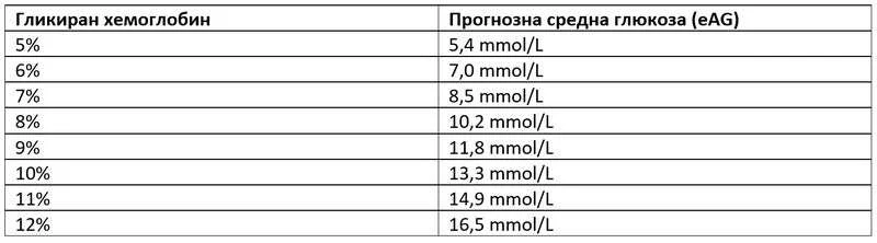
The accuracy of HbA1c depends on the lifespan of red blood cells. Conditions such as anemia, kidney failure, or bleeding can falsely lower or raise it.
How is insulin resistance testing done?
The most accurate method for assessing insulin sensitivity is the so-called. hyperinsulinemic euglycemic clamp (HEC) – the „gold standard“ that directly measures glucose absorption. However, this test is only used in research centers.
In practice, we rely on tests that indirectly assess insulin resistance (IR) by measuring glucose tolerance.
Oral glucose tolerance test (OGTT) with 75 g glucose is the most informative routine method. It shows the response of the pancreas and tissues to real load, which makes it more accurate than single fasting measurements. OGTT norms:
- KZ 0min: below 5.5 mmol/l (5.6−6.9 is prediabetes – a sign of IR)
- BG 60min: below 11 mmol/l (above 11 is diabetes)
- KZ 120min: below 7.8 mmol/l (over 7.8 is IR)
OGTT can be done as needed and more frequently than once a year, for example every 3–6 months if there is a change in weight or diet. There is no risk of „pancreatic exhaustion.“.
Fasting glucose and insulin measurements remain useful, especially for calculating the HOMA-IR and QUICKI indices. [35].
- HOMA-IR: The optimal value is 1, and value over 2 talk about IR.
- QUICK: The norm is 0.35−0.45. Value below 0.339 is IR.
- Fasting glucose above 5.5 mmol/L also indicates prediabetes and indirectly IR.
Insulin measurement has limitations (different methodologies, variability between laboratories). However, studies suggest:
- 2.5–10 μIU/ml is normal.
- 10 and 18 μIU/ml indicate possible IR.
- Above 18 μIU/ml – IR is very likely.
- During OGTT: Insulin above 150 for the first hour and above 60 for the second hour is also indicative of IR.
Tests such as OGTT, CZ, and insulin are strongly influenced by the days before the test.
What is the preparation for an insulin resistance test?
Before undergoing an IR test, the patient must be aware of several factors that may distort the test results, and must report them to the selected specialist.
Factors such as prolonged fasting, ketogenic diet, severe illness, or alcohol abuse can worsen results and create a false picture of impaired tolerance. [36].
It is recommended that you have at least 3 days of normal carbohydrate intake (about 150 g/day) before the test. If you fall into a risk category (starvation, eating disorders, etc.), be sure to notify your doctor.
If you think you may have IR or have already been diagnosed with it, the most sensible step is to consult an experienced specialist, with whose help to start effective diet therapy.
The most successful approach to addressing the problem is diet therapy, when it is individualized, balanced, and sustainable for long-term compliance.
If you found this article helpful, feel free to follow me on social media for more evidence-based content.
